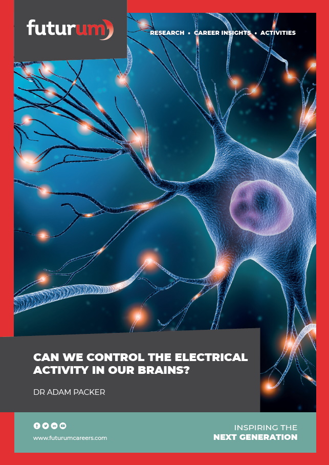
Unlocking new neuroscience frontiers by imaging the intricacies of the mouse brain
[[{“value”:”
Unlocking new neuroscience frontiers by imaging the intricacies of the mouse brain
Many mysteries of the brain remain unsolved. At Duke University in the US, Professor Allan Johnson and Associate Professor Leonard White are pushing the boundaries of neuroscience by imaging mouse brains to the highest-ever level of detail. Their Duke Mouse Brain Atlas will help neuroscientists around the world reveal more secrets about the brain.
Talk like a neuroscientist
Alzheimer’s disease — a neurodegenerative disease that causes dementia (symptoms include deterioration of memory, cognition and behaviour)
Antibody — a protein produced by the immune system that can be engineered to attach to other target proteins
Fluorescence — the absorption of electromagnetic radiation of one wavelength that results in re-emission of radiation at a different wavelength
Histology — the study of cell and tissue anatomy at the microscopic level
Neurodegenerative disease — a progressive loss of neurons leading to impaired brain function
New and emerging technologies are rapidly accelerating the pace of neuroscience research. At Duke University, Professor Allan Johnson and Associate Professor Leonard White are on the crest of this wave of innovation. They are merging three cutting-edge imaging techniques to build the world’s most comprehensive and detailed model of the mouse brain, which will act as a reference tool for neuroscience researchers and students around the world.
Imaging technique 1: magnetic resonance histology
Magnetic resonance imaging (MRI) is a diagnostic medical technique that creates detailed images of structures within the body. These images are created by placing the patient or specimen in a strong magnetic field that causes protons to oscillate (wobble) at a very specific radio frequency. Radio waves at these frequencies excite the protons, which return a signal that can be used to map the molecular environment of the tissue. Al, Len and their team have developed an incredibly powerful MRI machine to study the mouse brain at the cellular level. “Histology is normally performed using a microscope,” explains Al. “With magnetic resonance histology (MRH), we replaced that conventional optical microscope with an MRI microscope to observe the structure of tissues in the brain.”
The team’s machine operates on a much finer scale than clinical MRI. “Our MRH images are up to 2.4 million times more detailed than medical MRI images,” says Al. “To get this level of detail, physicists and engineers in our team spent over 40 years building a unique scanner to provide the most detailed images of the mouse brain ever acquired. The scanner employs a magnet that is up to 6 times stronger than most clinical machines.”
Imaging technique 2: light sheet microscopy
Len and Al complement their MRH data with light sheet microscopy (LSM) images. LSM involves shaping a laser beam into a thin sheet of light to create many two-dimensional images that are reconstructed into a three-dimensional representation of the whole sample. “First, we remove any components of the biological sample that scatter light – in other words, we make the mouse brain completely transparent,” says Al. “This involves soaking it in a collection of clearing chemicals.”
Next, fluorescent antibodies are added, which bind to specific proteins in the brain tissue. “When excited by certain wavelengths of radiation, the antibodies fluoresce,” explains Len. “This means we can see exactly where they – and the proteins they’re bound to – are in the sample.” Different neurons in the brain have distinct proteins, which means they can be targeted and labelled by antibodies at a very fine level. “The same method can also be used to show non-cellular proteins, such as the beta-amyloid protein associated with Alzheimer’s disease,” says Len. The resultant LSM images are at sufficient resolution to reveal the shape and structure of individual neurons in the mouse brain.
Imaging technique 3: micro-computed tomography
MRH and LSM images have some limitations: “When a mouse brain is removed from its skull, it becomes distorted and there are no longer any external landmarks for reference,” explains Al. To address these challenges, the team turned to micro-computed tomography (micro-CT), an imaging technique that takes multiple x-rays from different orientations and combines them to create a three-dimensional digital representation of the mouse skull.
By digitally placing their MRH and LSM mouse brain images inside their micro-CT model of the mouse skull, Al and Len can correct the distortion of the LSM images and provide external references from bony landmarks on the skull. The superb contrast in the MRH images allowed them to label 358 different internal brain structures. “Neuroscientists depend on such labels to share observations,” says Al. The result is the Duke Mouse Brain Atlas (DMBA) – a highly detailed three-dimensional representation of the tissues, cells, circuits and connections that together make up the mouse brain.
Insights and applications
Neuroscientists around the world can use the DMBA as a reference tool in their research as it allows them to locate different brain regions and reference changes in mouse models of disease and ageing. “For instance, we are using the DMBA to explore age-related neurodegenerative diseases,” says Len. “We’ve scanned over 100 brains of mice of different ages to assess how different regions of the brain change with age. We’ve also scanned nearly 500 brains of mice with genes linked to Alzheimer’s disease and mapped these onto the DMBA to observe how the brain changes as the disease progresses.”
The potential applications of the DMBA are near-endless. The exquisite brain images can be used as a teaching tool to inspire the next generation of neuroscientists. For example, the NeuroTok Initiative is engaging students with the DMBA by encouraging them to create fun and informative educational videos for social media that explain the complex structure of the brain. Researchers can use the DMBA to implant electrodes or probes into specific regions of living brains more accurately. MRI images can be labelled with more confidence. And neuroscientists will be able to answer questions about the relationship between brain structures (seen in MRH images) and specific neurons (seen in LSM images). “Together, these advantages will lead to better science and more precise and replicable results,” says Al.
Thanks to the creation of the Duke Mouse Brain Atlas, Len and Al have made the beauty of the brain visible, enabling everyone to appreciate the wonders of this mysterious organ.
 Professor G. Allan Johnson
Director, Duke Center for In Vivo Microscopy
Departments of Radiology, Physics and Biomedical Engineering, Duke University, USA
Fields of research: Radiology, physics, biomedical engineering, neuroscience
Associate Professor Leonard E. White
Associate Director, Duke Institute for Brain Sciences
Department of Neurology, Duke University, USA
Fields of research: Neuroanatomy, neurophysiology, brain development and evolution, medical education
Research project: Using cutting-edge imaging technologies to build the Duke Mouse Brain Atlas
Funders: US National Institutes of Health (NIH): National Institute of Aging (NIA, grant R01 AG070913-01); National Institute of Neurological Disorders and Stroke (NINDS, grant R01 NS120954-01A1)
Reference
https://doi.org/10.33424/FUTURUM620


About neuroscience
Neuroscience is the scientific study of the nervous system, in particular the brain. “Embracing the challenge of understanding the brain is an exciting adventure,” says Len. “As technology advances and more young people get involved in this pursuit, we are sure to discover even more.”
Impactful neuroscience research involves combining expertise from many disciplines, as the Duke Mouse Brain Atlas project demonstrates. Al uses his physics and biomedical engineering background to help develop the sophisticated equipment needed, while Len brings his in-depth knowledge of mammalian brain anatomy. “I also bring my knowledge and experience in medical neuroscience to help understand how our work can be best translated into real-world applications,” says Len.
Len and Al have now been working together for over fifteen years. “The science we have pursued could not have been accomplished without our mutual willingness to learn from, with, and about the disciplines we represent,” says Len. “Such collaborations are crucial for progress and innovation in neuroscience. The Duke Mouse Brain Atlas would not have been possible without such interdisciplinary collaboration – science is a team sport and the best results occur when we work together.”
The rate and breadth of discoveries within neuroscience continue to increase exponentially. “I hope that the next generation will build understanding about how brain circuits give rise to consciousness,” says Len. “I also hope that we learn more about how to shape the brain’s plastic potential (ability to adapt) to help people flourish, whether that means treating or resisting disease or simply being the best versions of themselves.”
Pathway from school to neuroscience
At high school, study biology, physics, chemistry, mathematics and computer science to get a foundation in the knowledge and skills required for studying neuroscience.
“Invest in the quantitative sciences and value the breadth of the biological sciences,” advises Len. “Wonder with awe and inspiration at the amazing diversity of life in all its forms. And appreciate the richness of human experience expressed through the humanities and arts.”
At university, you could study a degree in neuroscience. However, as neuroscience is such an interdisciplinary field, it requires people with different areas of expertise. You could also study a degree in biology, psychology, chemistry, biomedical engineering, medicine or computer science and apply your skills in neuroscience.
Explore careers in neuroscience
The Duke Institute for Brain Sciences organises the Duke University Neuroscience Experience (DUNE), a summer research programme for local high school students.
Duke University also hosts wider outreach programmes that include opportunities to explore neuroscience, including the Duke Research in Engineering Program, the Duke Cell Biology Academy, Building Opportunities and Overtures in Science and Technology and the Duke Health Professions Recruitment and Exposure Program.
Meet Al

I have been interested in physics from my earliest childhood. When I was younger, I read Scientific American avidly. In seventh grade, I made a model of an oxygen atom and declared to my parents that one day I would become a university physics professor!
As a teenager, I was into music as well as science. I played the clarinet and saxophone in my high school band, sang in my church choir, and played the banjo in a folk trio which was a big hit at the local Rotary.
I finished graduate school in 1974, when CT and MRI technologies were just emerging. I was fortunate beyond my wildest dreams to get a job in the Department of Radiology at Duke University at the right place at the right time. I joined the Biomedical Engineering faculty in 1994 where I have mentored some truly extraordinary undergraduate, graduate and medical students.
I love the beauty of the brain’s anatomy. The exquisite detail of our mouse brain images is glorious! I enjoy loading the DMBA into sophisticated software that lets us view the brain in many dimensions. With Len as our guide, we explore the connections between the different structures of the brain.
I enjoy spending my free time in my wonderful workshop in the woods where I make furniture and turn bowls. I am currently learning to use a new computer-controlled cutting machine from my son’s company, which involves quite a steep learning curve!
Al’s top tips
Read as much as you can, walk in the woods and give yourself space to think about what could be.
Meet Len

As a teenager, I was interested in music and spending time in the natural world. I grew up playing the clarinet and saxophone but have since realised that my musical soul is best expressed through the acoustic guitar.
I came from a lower-income background. I knew nothing about scientific careers, and my brother was the only person in my family who had graduated from university. I went to university to study biology and marine ecology, where I discovered my love of learning about life and how it works.
I pursued graduate studies in biomedical science because I had become interested in cardiovascular physiology. But one fateful day, a faculty member told me: “The future is not in the heart, it’s in the brain.” I took this advice to heart (pun intended!) and decided to check out the neuroscience labs at the other end of the hall. There, I fell in love with the brain. More than four decades later, I remain in wonder and humble fascination with the beauty and mysteries of the brain.
I love to think about how no two brains are identical. Like faces and fingerprints, each is distinctively unique in its size, pattern and complexity.
In my free time, I enjoy spending time with my family. I’m an avid runner, keen tennis player and occasional pickleball player, and I enjoy watching college sports. My classical guitar skills are reaching advanced amateur level, I am a faithful member of a local congregation, and I enjoy hiking and backpacking adventures with my wife.
Len’s top tips
Cultivate curiosity. Make time for reflection and allow your mind to wander creatively. This is how the best questions are formed, which lead to new discoveries, insights and understanding. These questions may be the start of your greatest adventure.
Do you have a question for Al and Len?
Write it in the comments box below and Al and Len will get back to you. (Remember, researchers are very busy people, so you may have to wait a few days.)

Learn about the types of neuroscience research that will benefit from the Duke Mouse Brain Atlas:
futurumcareers.com/can-we-control-the-electrical-activity-in-our-brains
The post Unlocking new neuroscience frontiers by imaging the intricacies of the mouse brain appeared first on Futurum.
“}]]


